The Clarkson University Orthopaedic Biomechanics Laboratory is now closed.
From 2009 to 2024, the lab trained 9 graduate and 35 undergraduate researchers. We pushed the boundaries of orthopaedic research using the finest locally-harvested materials and advanced biomedical engineering knowledge, specifically about biomechanics. Lab alumni (including Dr. K.) now hold positions in academia, industry, and government.
This site is now archival as of 6/15/2024.
Biomechanics, a type of biomedical engineering, applies engineering principles to biological applications to reveal relationships between structure, function, and injury.
Take a look at our research below!
Injury Biomechanics
Vertebral Fractures:

Vertebral fractures can be debilitating and can occur due to traumatic injury or due to the cumulative effects of repetitive loading. Using a cervine ex-vivo model, our work has shown that fractures of the vertebral ring apophysis can occur under repetitive loading in the absence of traumatic loading in otherwise-healthy lumbar vertebrae. This work will ultimately lead to injury prevention recommendations for those who participate in high-cycle low-load activities, such as sports.
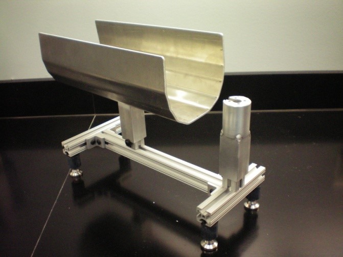
Meniscal Injuries:
Meniscal injuries occur in active populations by way of a compressive and torsional load and are characterized by a fibrous rupture in the meniscus. When evaluating current repair methods, meniscal injuries are simulated by scalpel incisions; however, these incisions are poorly representative as they lack fibrous rupture. This work intends to create a novel method to simulate meniscal injuries by using large compressive forces. Ultimately this work aims to improve how meniscal repair methods can be tested.
Orthopaedic Device Design
Adjustable-length Intramedullary Nail:
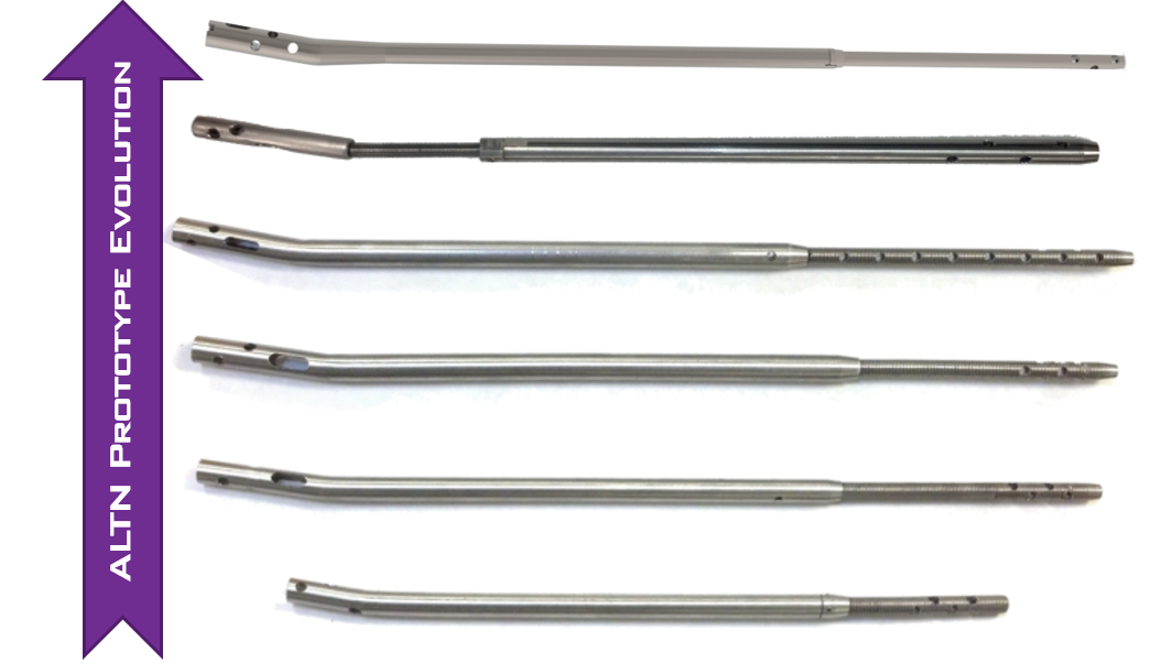
View Here. Intramedullary (IM) nails are used to reduce approximately 50% of all tibia fractures in the United States. For optimal healing, the IM nail must be an appropriate length for the patient, otherwise complications such as periprosthetic fractures or nonunion can result. An adjustable length IM nail would reduce manufacturing cost, inventory cost, and patient complications. The patented design of our prototypes matched or surpassed the reported performance of discretely sized nails when tested in cervine and cadaver tibiae. Work on this project supported the creation of Adaptable Ortho Innovations LLC, a small business determined to create adaptable medical devices for all patient anatomy. Our preliminary market and cost analysis suggests that the most eager customers will be smaller hospitals and trauma centers with limited cash flow in the US or in developing countries. The adjustable IM nail will benefit patients, the manufacturers, and orthopaedic providers.
Limited Motion Wrist Brace:
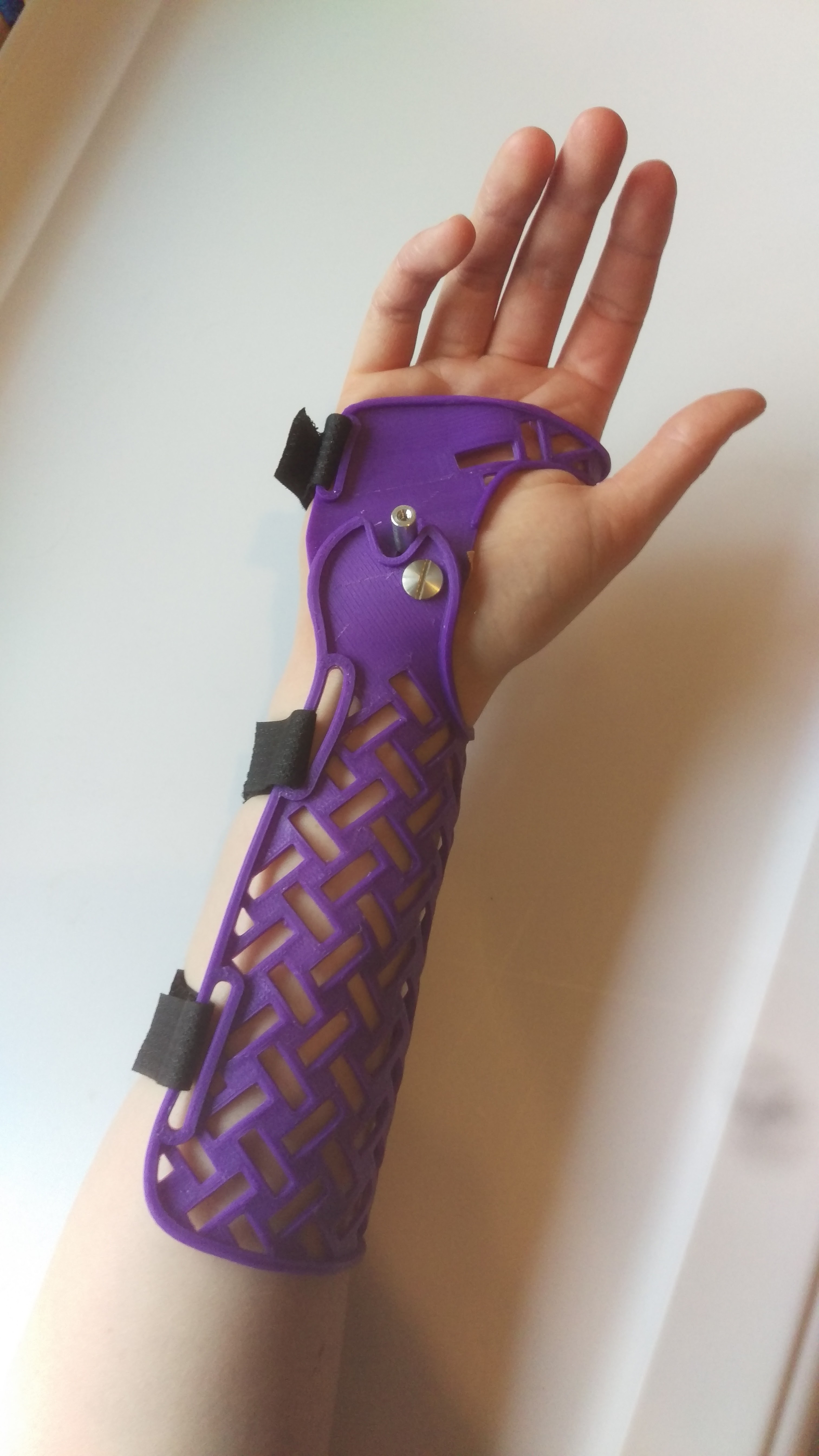
The scapholunate interosseous ligament is commonly injured, requiring surgical repair and subsequent immobilization. Limited motion can decrease healing times and increase patient compliance. There are few commercially available options for limited motion braces that permit safe movement in the Dart Thrower’s Plane (DTP). Our DART brace is custom manufactured by 3D printing using the polymer PETG and is custom fitted to the subject. Ongoing work is validating the motion limits of the design and assessing functional abilities of individuals while wearing the brace.
Cervical Collar:
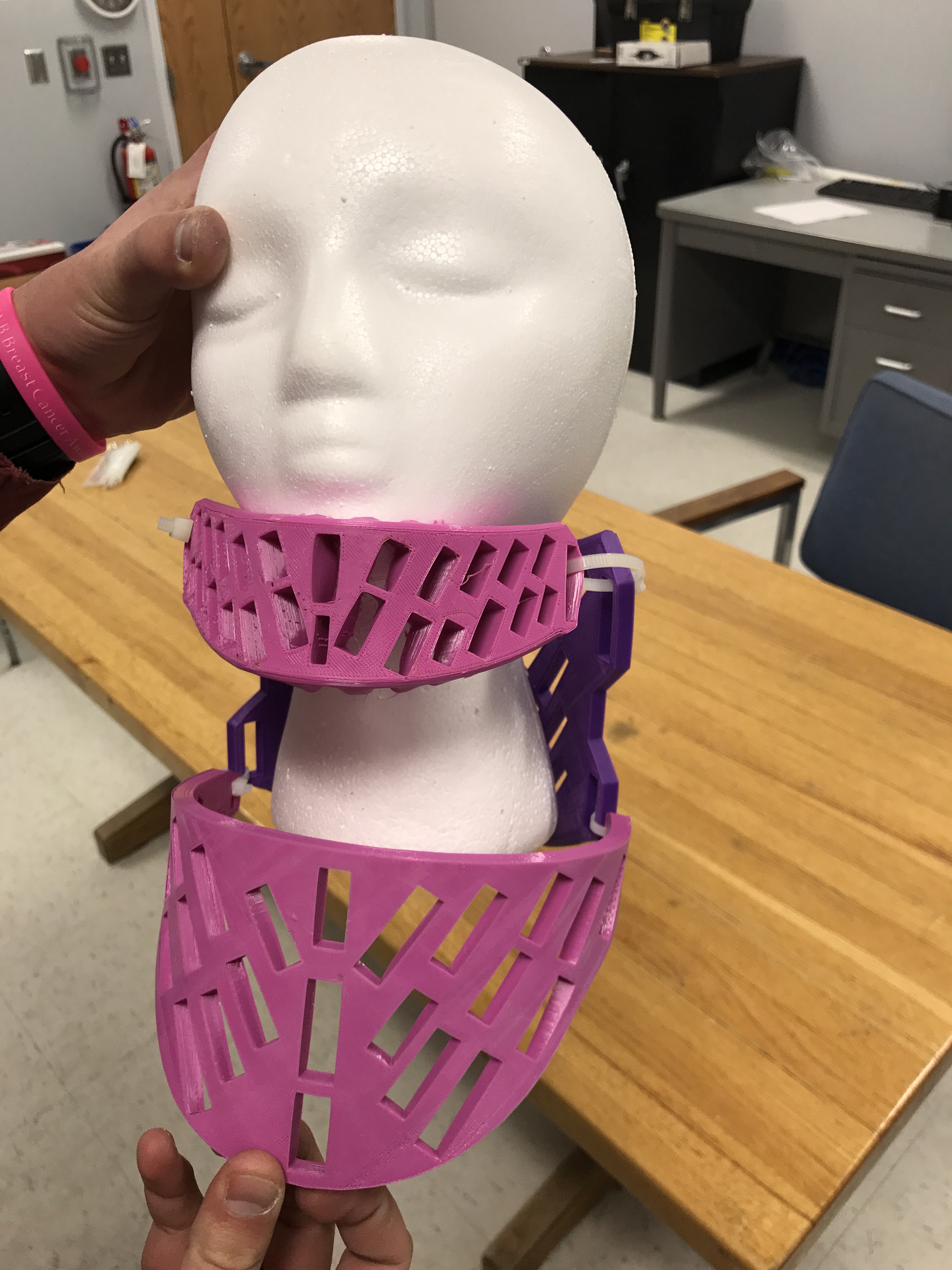
Cervical spine injuries are the most common types of spinal cord injuries. The upcoming design for a new cervical collar will be custom fit to the patient. The brace will be 3D printed then thermo-molded to the patient to provide the most comfort and support. There will be the option to change heights of the chin piece by a clinician, to alter the height and increase the motion of the neck to decrease physical therapy time. The rest of the brace will be held together by velcro. This brace is intended for someone who will need to be in a static brace after surgery or a server injury for long period of time.
Quantifying Upper Extremity Function
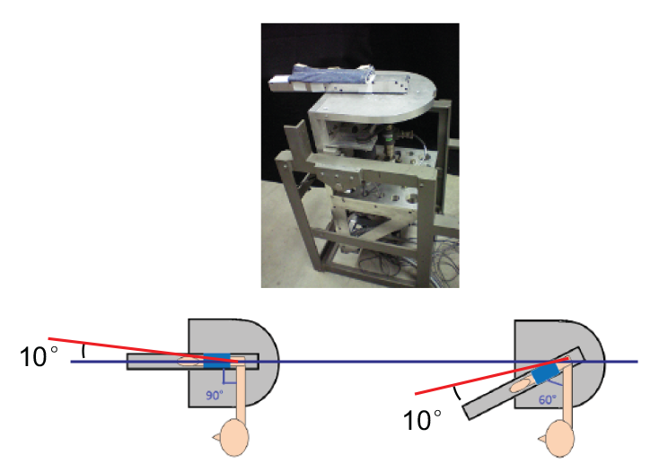
3D Hand Grip Forces:
Weak grip strength has been a correlating factor in predicting numerous health issues including heart disease, Type 2 Diabetes, and osteoporosis. The Jamar Hydraulic Hand Dynamometer, bulb dynamometers, and pinch dynamometers are all well-known methods in determining the muscular forces within the hand; however, these methods output the acting force vectors within the hand as a one-dimensional magnitude. The long-term goal of this research is to map and analyze the three-dimensional grip forces created by the hand in the standardized grip position to ultimately permit a greater specification for predictions of health ailments and quantify why these relationships exist, particularly in osteoporotic populations. Towards that long-term goal, the immediate goal is to measure three-dimensional (3D) hand grip forces and their fatigue rates in healthy volunteers.
Select Previous Research
Ex-vivo Knee Model:
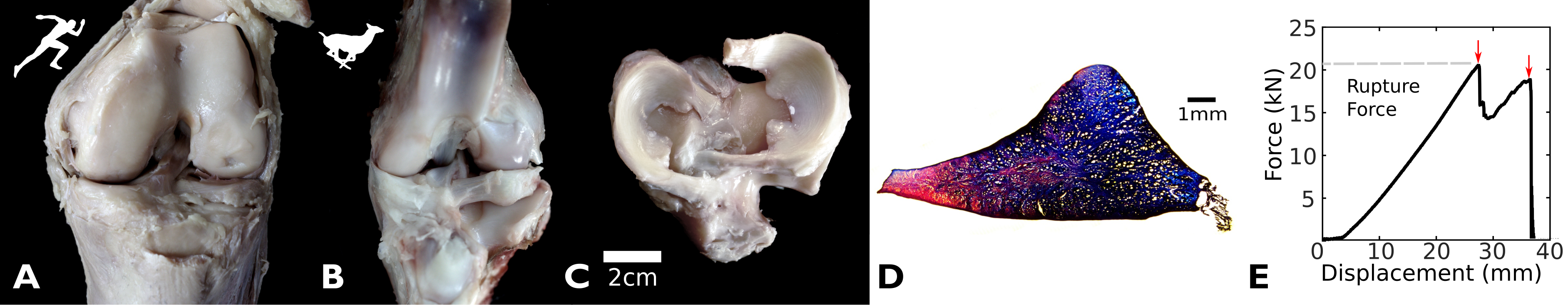
Animal models are valuable proxies for biomechanical studies. Accurate knee models assist surgeons in practicing new reconstruction techniques and are crucial to the advancement of technology and rehabilitation. Current quadruped knee models greatly differ from human knees in both aspects of size and morphometry. The cervine knee was evaluated as a novel animal knee model. We measured its range of motion, cross-sectional area and rupture strength of the anterior cruciate ligaments (ACL), bony morphometry, and compressive properties of the meniscus. This improved ex-vivo model will offer a testing platform for orthopaedic implants, a training analogue for surgeons, and will accelerate the translation of results from the laboratory to the clinic.
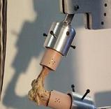
Elbow Joint Stiffness:
Elbow joint stiffness is an important clinical problem, affecting those who have certain diseases (such as Parkinson’s Disease) and those who suffer from traumatic elbow injuries. Clinically, quantifying elbow stiffness remains difficult at best. A device is under development to offer a quantification of elbow stiffness that will ultimately be used to physical therapists for monitoring changes in elbow stiffness during recovery. Additional applications to users of prosthetic limbs are possible.
Ligament Modeling:
Our understanding of the mechanical behavior of biologic tissues is constantly evolving. Accurately describing the stress-strain relationship for ligaments is challenging, particularly for the medial ulnar collateral ligament (mUCL) of the elbow. This ligament has a unique two-band structure, such that one band is always in tension. The researchers are seeking a better way to mathematically describe the ligament and will validate the model with experimental tests of ligament behavior.
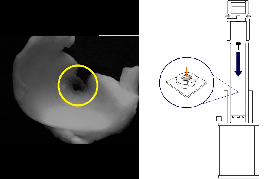
Bone Nanoindentation:
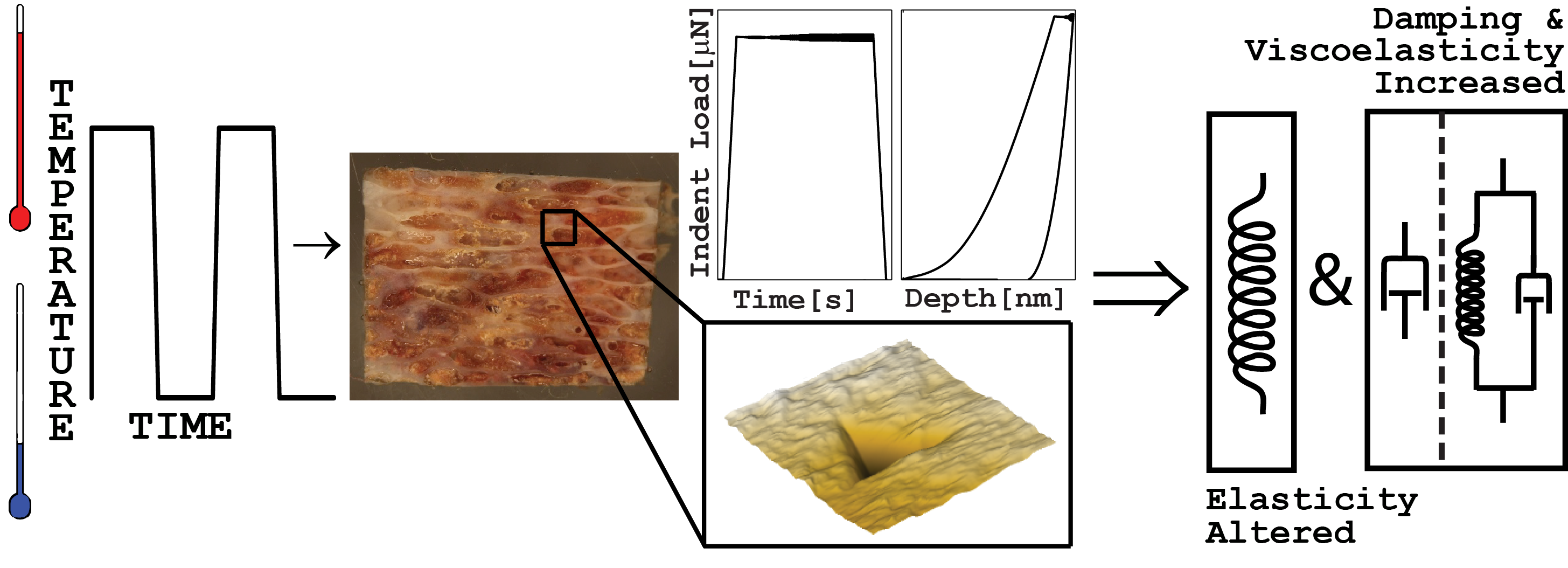
The properties of small-scale structures that make up the microstructure of bone are measurable via nanoindentation. Nanoindentation is an advanced material characterization technique in which a probe on the scale of hundreds of nanometers is forced into the surface of the material, while applied load and displacement are measured. This technique is being employed to determine the effects of freezing and thawing cycles on the material properties of bone, knowledge that will be applied to testing methods.
Other Services
Data Analysis and Manuscript Preparation for Clinical Studies:
Students and faculty from the Orthopaedic Biomechanics Laboratory have experience with data collection, analysis, and preparation for publication of clinical studies in orthopaedics. Contact us to discuss.