Pattern
formation in E.coli: a model for the localization of the division
site
by
the
pole-to-pole oscillations of Min proteins
- an overview
Formation
of stable patterns by local autocatalysis and depletion of a long-ranging
substrate
A minimum
mechanism to generate oscillating polar
patterns
Oscillation
in counter-phase and multiple division sites in long extended
filaments
Some
examples of the changing behavior after parameter
changes
Simulation
using a tube-like
geometry
A short
program that allows the simulation of the MinD/MinE waves
Time-lapse
fluorescence micrographs
Back to main entrance page:
Theoretical
aspects of pattern formation and neuronal development
To facilitate an
understanding of this highly complex process, first an analogy should be given
(knowing that all analogies are a bit dangerous). Imagine a strip of very
rapidly growing grass on which a cow is grazing. After eating up all grass in
the immediate surrounding, the cow will start to move into a region in which
more grass is available. In which direction to go first is more ore less random
but it will continue in this direction since less grass is left behind. After
reaching the end of the strip, the cow will move rapidly to the other side of
the strip where still fresh grass is available. After reaching the other end,
the process will start anew, assuming that the grass recovered meanwhile. In the
bacterium, a protein called MinD covers uniformly the membrane (the grass).
According to our model, a local signal (MinE, the cow) needs MinD to bind to the
membrane but removes thereby MinD from the membrane. Thus, the MinE signal will
move into a region where more MinD is available, and so on. In this way, the
minE signal sweeps over the field like a windshield wiper of a car. On time
average, the MinD concentration is lowest in the center, allowing the initiation
of the division apparatus (FtsZ-ring). In the analogy, the cow is anyway small
and localized in relation to the strip. To achieve a similar localization of a
biochemical reaction, a pattern forming reaction is required. By mathematical
modelling, the type of interactions are explored that account for the observed
dynamic behaviour of the substances involved.
More scientifically
speaking, the preparation for division starts with the assembly of a polymeric
ring of the tubulin-like GTPase FtsZ (Z-ring). In E.coli, this ring is
localized to the center by the actions of the MinC, MinD, and MinE proteins.
MinC inhibits the initiation of the Z-ring. MinC co-localizes with MinD. In
wildtype cells (WT), MinC/D forms a polar pattern that oscillates between the
poles, keeping the center free for initiation of cell division. Thus, virtually
all of MinC/D dynamically assembles on the membrane in the shape of a test-tube
covering the membrane from one pole up to approximately midcell (time-lapse
fluorescence micrographs). Most of MinE accumulates at the rim of this tube,
in the shape of a ring (the E-ring). The rim of the MinC/D tube and associated
E-ring move from a central position to the cell pole until both the tube and
ring vanish. Meanwhile, a new MinC/D tube and associated E-ring form in the
opposite cell half and the process repeats, resulting in a pole-to-pole
oscillation cycle of the division inhibitor. A full cycle takes about 50s. The
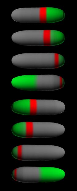
panel below shows a schematic drawing of the MinC/D (green) and MinE (red)
localization cycles. The animations show a typical computer simulation using our
model to describe the dynamic behaviour of these proteins (Meinhardt
and de Boer,
2001).
The following simulations
illustrate the elementary
steps:
Formation
of stable patterns by local autocatalysis and depletion of a long-ranging
substrate
A minimum
mechanism to generate oscillating polar
patterns
Oscillation
in counter-phase and multiple division sites in long extended
filaments
Some
examples for the changing behavior after parameter changes
Simulation
using a tube-like
geometry
A short
program that allows the simulation of the MinD/MinE waves
Last
update:
ralf.dahm@tuebingen.mpg.de