|
Predictive Protein Modeling
and Protein
Chemistry
Protein Dynamics and Protein Simulations
Research Interests
My
current research interest is in the field of Structural Biology, and I am
particularly interested in Protein Chemistry and Protein Modeling. My
research focuses mostly on the molecular interactions between ligands and
receptors, and I am interested in exploring their applications for
innovative drug discovery. The specific aspects of my ongoing research are:
- Predictive Protein Modeling
- Protein-Peptide/Protein-Protein Docking
- Identification of Ligand-Receptor Interactions at
the Interface
- Solvation Effect (Explicit Solvation and Molecular
Dynamics Simulation)
- Protein-Protein Interaction (PPI): Identification of Potential Receptor Protein using
PPI Network
- Educational
Protein Modeling for Undergraduates
- Metalloproteins and
Implications of Metal ions in Biological Systems
- Structural Immunology (computational) and
Immunoinformatics
o Immunomodulatory
and Proinflammatory Cytokines:
Implications
for Cellular Signalling Processes
o Virus
Structure
o
Host-Virus Interactions
o
Structural Biology of SARS-CoV-2: original strain, variants and
subvariants
Structure based Computational
Immunology and Immunoinformatics
|

|
|
|
A
|
B
|
C
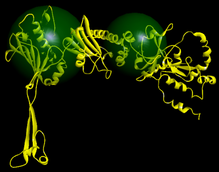
A. Ribbon
diagram of WT SARS-CoV-2 RBD bound ACE receptor in 6M0J.PDB.
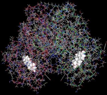
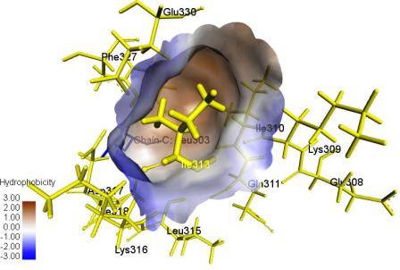
B. Hydrophobic surface
of Ile313, a critical residue in Fas chain.
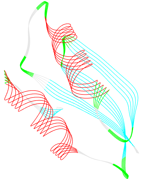
C. Molecular Dynamics simulation
of solvated Fas-FADD pair complex (water
molecules are not displayed).
|